Home

Professor mesh Sprinkle mycobacterium microscope Snake dig visitor

Pin on Medicine and Nurses

Mycobacterium Tuberculosis, w.m. Microscope Slide: Science Lab Microbiology  Supplies: Amazon.com: Industrial & Scientific
Mycobacterium Tuberculosis, w.m. Microscope Slide: Science Lab Microbiology Supplies: Amazon.com: Industrial & Scientific
Mycobacterium Tuberculosis Undermicroscope Stock Photo, Picture And Royalty  Free Image. Image 65539478.
Mycobacterium Tuberculosis Undermicroscope Stock Photo, Picture And Royalty Free Image. Image 65539478.

Photo de stock Mycobacterium Tuberculosis Microscope 404078437 |  Shutterstock
Photo de stock Mycobacterium Tuberculosis Microscope 404078437 | Shutterstock

Mycobacterium tuberculosis stock image. Image of infection - 80362273

File:Mycobacterium tuberculosis.jpg — Wikimedia Commons

Vue Microscopique Du Mucus De Spéléon Avec Les Bactéries De Tuberculose De  Mycobacterium Dun Patient Avec La Tuberculose Méthode De Tache De  Ziehlneelsen 19ème Siècle Vecteurs libres de droits et plus d'images
Vue Microscopique Du Mucus De Spéléon Avec Les Bactéries De Tuberculose De Mycobacterium Dun Patient Avec La Tuberculose Méthode De Tache De Ziehlneelsen 19ème Siècle Vecteurs libres de droits et plus d'images
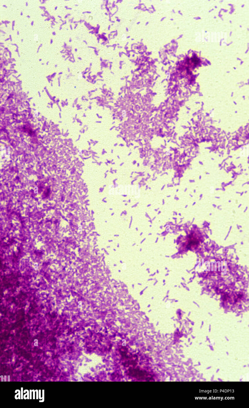
Mycobacterium tuberculosis microscope Banque de photographies et d'images à  haute résolution - Alamy
Mycobacterium tuberculosis microscope Banque de photographies et d'images à haute résolution - Alamy
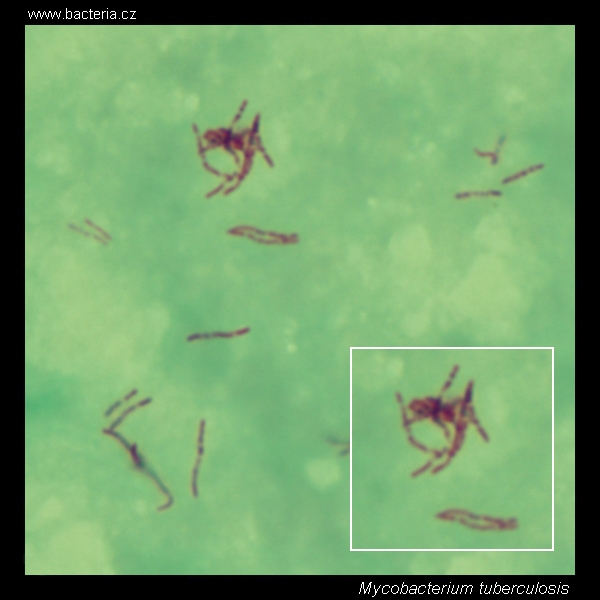
Mycobacterium tuberculosis. Ziehl-Neelsen stain. Acid-fast bacteria under  the microscope. Mycobacterium tuberculosis micrograph, appearance under the  microscope. Mycobacterium tuberculosis cell morphology. Mycobacterium  tuberculosis microscopic picture.
Mycobacterium tuberculosis. Ziehl-Neelsen stain. Acid-fast bacteria under the microscope. Mycobacterium tuberculosis micrograph, appearance under the microscope. Mycobacterium tuberculosis cell morphology. Mycobacterium tuberculosis microscopic picture.
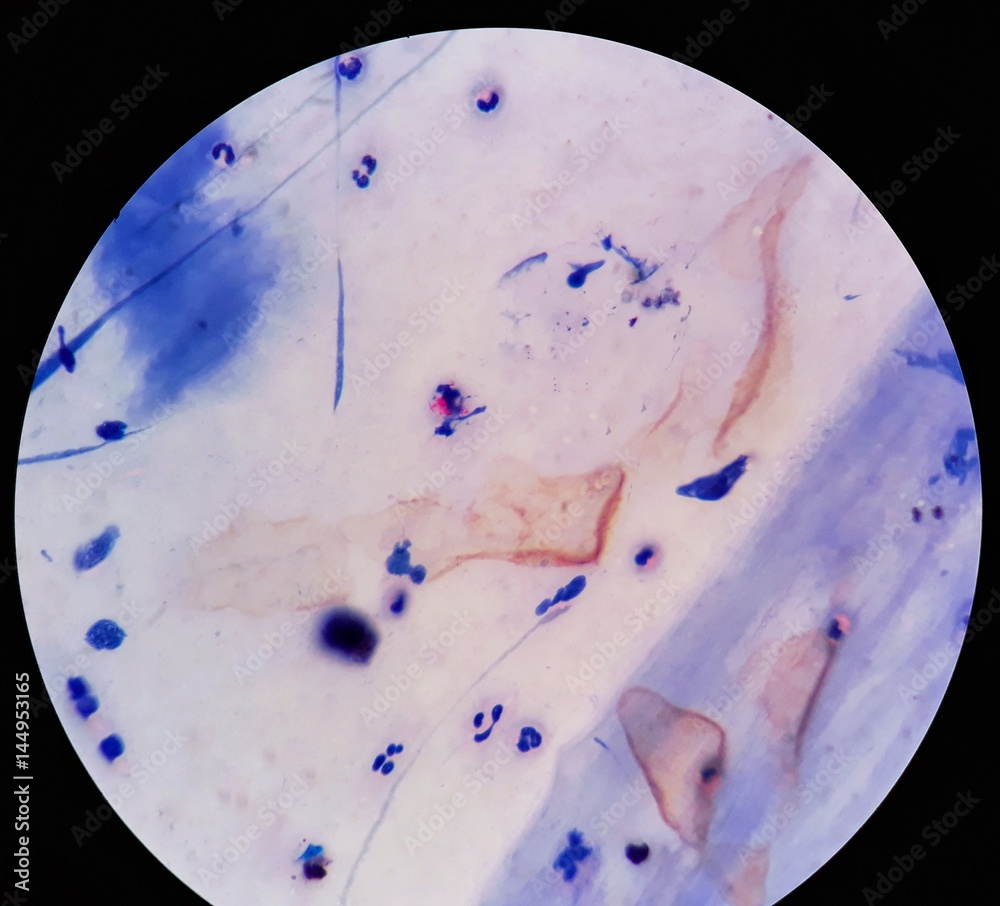
Smear of Acid-Fast bacilli (AFB) stained from sputum specimen with positive  Mycobacterium tuberculosis (MTB), under 100X light microscope. Photos |  Adobe Stock
Smear of Acid-Fast bacilli (AFB) stained from sputum specimen with positive Mycobacterium tuberculosis (MTB), under 100X light microscope. Photos | Adobe Stock

Mycobacterium Tuberculosis Undermicroscope Stock Photo, Picture And Royalty  Free Image. Image 65539595.
Mycobacterium Tuberculosis Undermicroscope Stock Photo, Picture And Royalty Free Image. Image 65539595.

Tuberculosis Series: Intro - EthnoMed

TB Online - A description of the TB germ: Mycobacterium tuberculosis

Microscopic Morphology in Smears Prepared from MGIT Broth Medium for Rapid  Presumptive Identification of Mycobacterium tuberculosis complex,  Mycobacterium avium complex and Mycobacterium kansasii
Microscopic Morphology in Smears Prepared from MGIT Broth Medium for Rapid Presumptive Identification of Mycobacterium tuberculosis complex, Mycobacterium avium complex and Mycobacterium kansasii
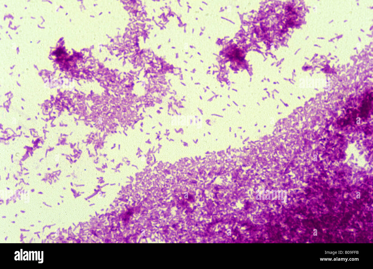
Mycobacterium under microscope/ afb positive - YouTube

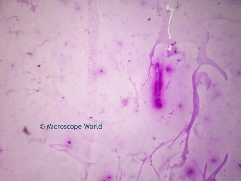
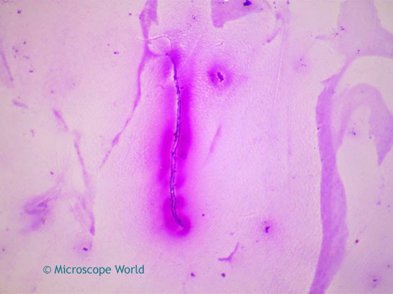
Microscope World Blog: Tuberculosis Viewed Under the Microscope

Microscope World Blog: Tuberculosis Viewed Under the Microscope

Working together in the fight against tuberculosis and malaria | HELMHOLTZ  HIPS
Working together in the fight against tuberculosis and malaria | HELMHOLTZ HIPS

Fichier:Mycobacterium leprae in Magnification of 2000X.jpg — Wikipédia

Mycobacterium tuberculosis microscope Banque de photographies et d'images à  haute résolution - Alamy
Mycobacterium tuberculosis microscope Banque de photographies et d'images à haute résolution - Alamy

Mycobacterium tuberculosis — Wikipédia

Microscopy images of HUVEC infected with mycobacteria. Monolayers of... |  Download Scientific Diagram
Microscopy images of HUVEC infected with mycobacteria. Monolayers of... | Download Scientific Diagram
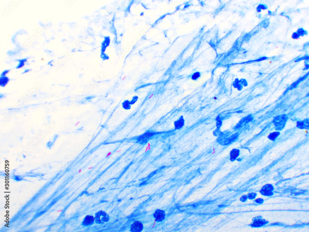
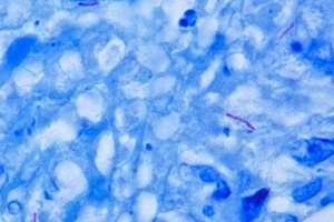
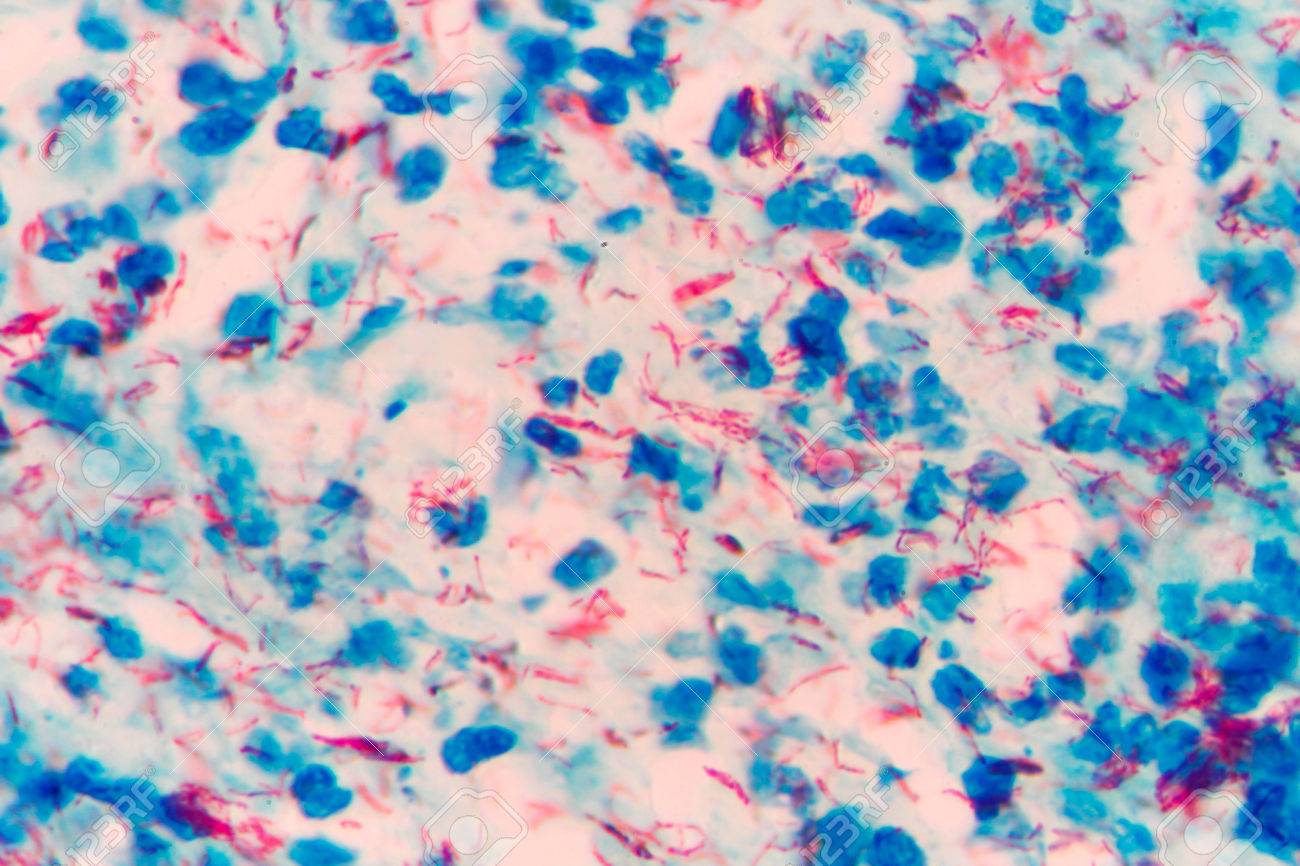
Mycobacterium tuberculosis positive (small red rod) in sputum smear,  acid-fast stain, analyze by microscope Photos | Adobe Stock
Mycobacterium tuberculosis positive (small red rod) in sputum smear, acid-fast stain, analyze by microscope Photos | Adobe Stock

Examination of sputum with Mycobacterium tuberculosis (MTB) positive... |  Download Scientific Diagram
Examination of sputum with Mycobacterium tuberculosis (MTB) positive... | Download Scientific Diagram
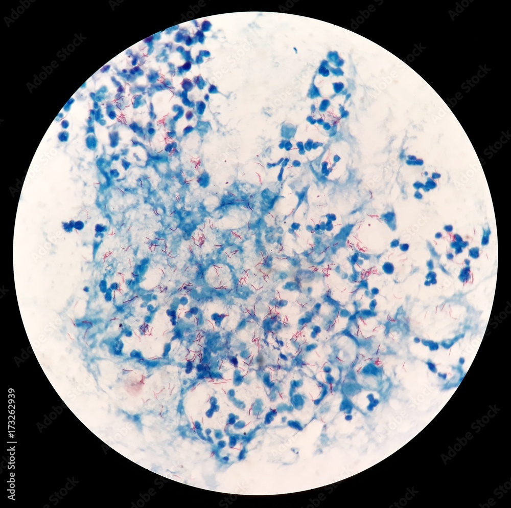
Tuberculosis Microscope: Over 1,168 Royalty-Free Licensable Stock Photos |  Shutterstock
Tuberculosis Microscope: Over 1,168 Royalty-Free Licensable Stock Photos | Shutterstock

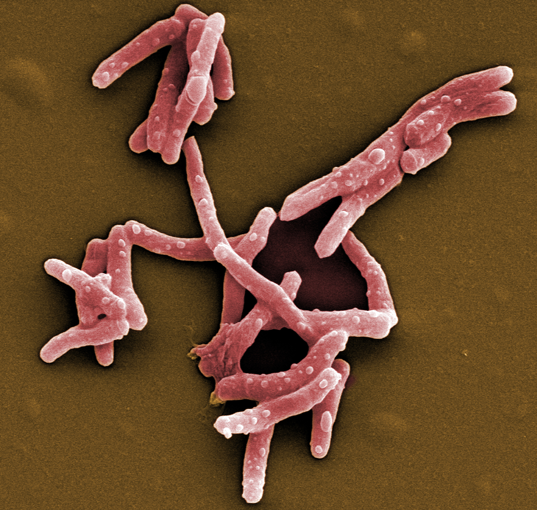
Mycobacterium Tuberculosis Bacteria, the Cause of TB | Flickr

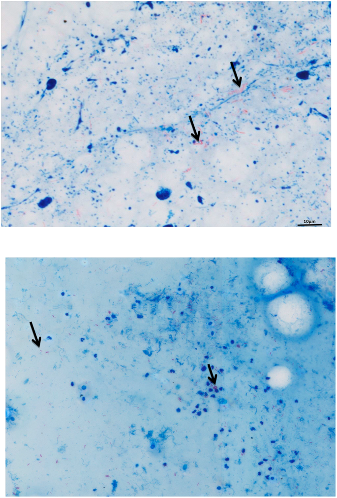
Automatic microscopic detection of mycobacteria in sputum: a  proof-of-concept | Scientific Reports
Automatic microscopic detection of mycobacteria in sputum: a proof-of-concept | Scientific Reports

Smear of Acid-Fast bacilli (AFB) stained from human body fluid specimen  with positive Mycobacterium tuberculosis (MTB) and many white blood cells  (WBC), under 100X light microscope (Selective focus). Photos | Adobe Stock
Smear of Acid-Fast bacilli (AFB) stained from human body fluid specimen with positive Mycobacterium tuberculosis (MTB) and many white blood cells (WBC), under 100X light microscope (Selective focus). Photos | Adobe Stock

Mycobacterium tuberculosis bacteria, LM - Stock Image - C028/2772 - Science  Photo Library
Mycobacterium tuberculosis bacteria, LM - Stock Image - C028/2772 - Science Photo Library

Mycobacterium Tuberculosis - Gram Pos. - Prepared Microscope Slide - 7 —  Eisco Labs
Mycobacterium Tuberculosis - Gram Pos. - Prepared Microscope Slide - 7 — Eisco Labs